Author: Cheyanne Durham
Date: August 16, 2022
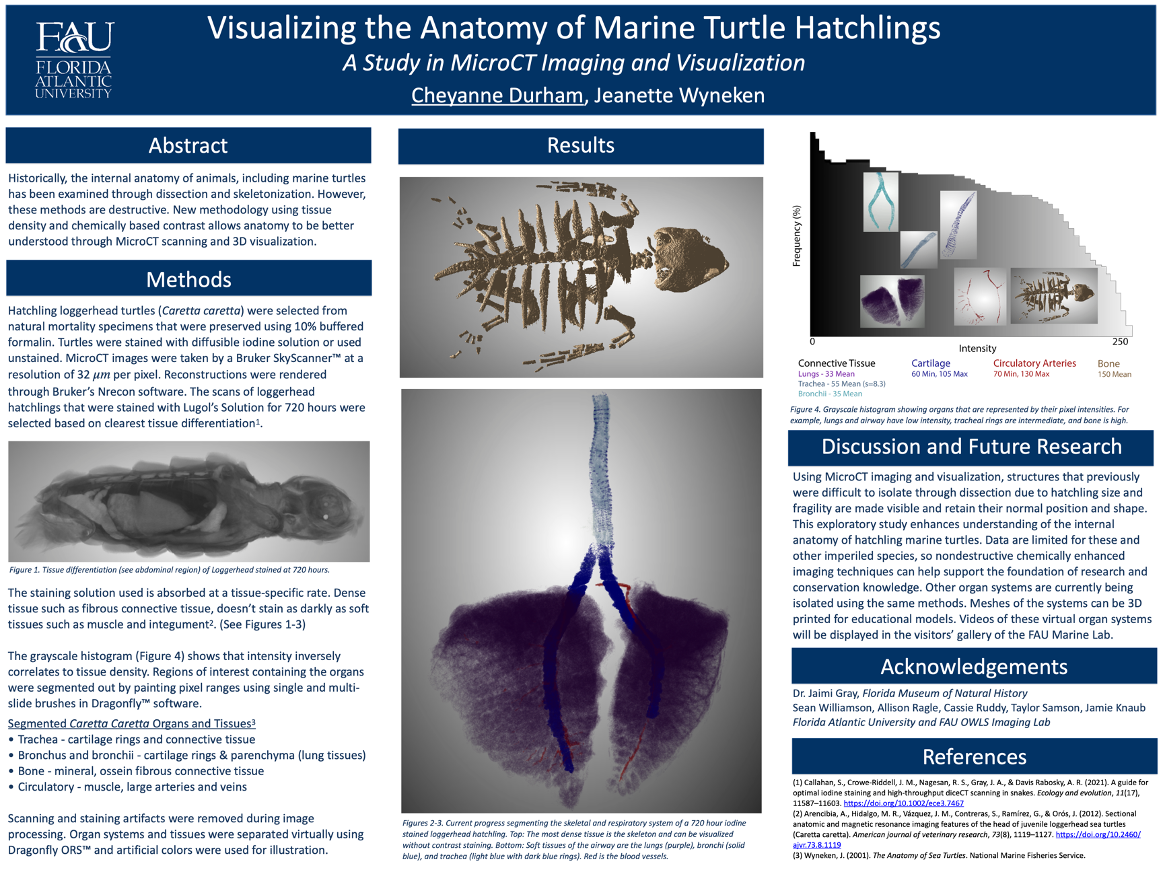
For the past eight months, the FAU Marine Science Lab has been “looking under the shell” to understand the anatomy of loggerhead hatchlings. Using soft tissue staining and MicroCT scanning, we can visualize the organs of naturally deceased hatchlings without destroying fragile, delicate structures. Ultimately, we are working towards creating a fully virtual 3D model of the internal anatomy of a hatchling sea turtle.
Staining
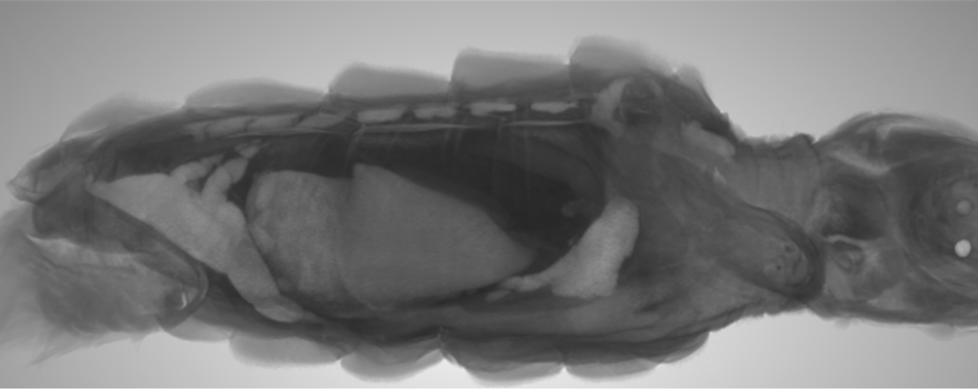
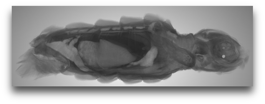
First, a hatchling loggerhead (preserved, dead from natural causes, and obtained with the proper permit) was stained with a dye called Lugol’s solution (potassium iodide). Both iodide and the cell membranes that make up the tissues within the turtle are negatively charged. Denser tissues repel more of the potassium iodide molecules, because they have more negative charge and therefore they stain lighter color. Less dense tissues have less negative charge, and therefore, retain more potassium iodide and stain darker.
Scanning
Thanks to our friends at the FAU OWLS Imaging Lab, we scanned the stained turtle using a MicroCT scanner. This scanner works just like a CAT scan that uses X-rays, but at a much smaller scale for our hatchling! Hundreds of pictures are taken at different angles to generate computerized cross-sectional images.
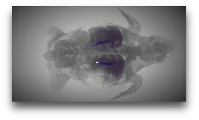
Segmenting
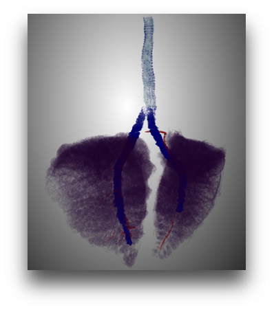
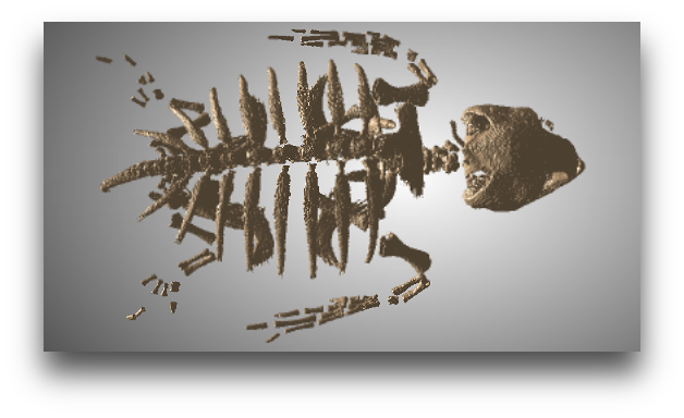
Using software for reconstructing these many pictures - Dragonfly ORS™ - we can see the cross sectional images of our scanned hatchling. The software combines the stacks of images to create a virtual 3D model. Using what we know about the chemistry of the dye and how tissues react to it (see staining above), we can separate out specific organs by their tissue density. Bone is denser than lung tissue, therefore, it shows up brighter on our scans. Going picture-by-picture, we use different computerized techniques to isolate out regions that contain the organs. See our progress on the respiratory and skeletal system!
In August, this research was presented at the American Chemical Society’s FAME (Florida Annual Meeting and Exposition) conference in Tampa. There, everyone asked questions to understand the big picture - why conduct such time intensive research on loggerhead turtle hatchlings?
(a) The loggerhead sea turtle is a threatened species in Florida and endangered elsewhere in the world. By sharing this knowledge, researchers and the public can gain an in-depth understanding of marine turtle anatomy without going through lengthy and sometimes restrictive permitting procedures. These virtual models are also printable in 3D, making them an asset in all teaching environments - especially because literature on the subject is limited.
(b) Sea turtles are an important indicator species. This means that they are sensitive to changes in their environment and their health
and population tell us much about the condition of our beaches and reefs. The more we understand their normal anatomy , the easier it is to recognize potential pathologies. The turtles can tell us the state of the environment we share with them.
(c) Similar computerized analyses of tissue density have been used to understand and treat human morbidities - such as osteoporosis and lung cancer. As the science progresses, with enough samples for statistical analysis, research like this could help guide rehabilitation and therapeutic approaches for marine turtles.
Want to know more about this research? Check out the poster from the ACS conference below and visit us at the Gumbo Limbo Environmental Complex.