Author: Maggie Dalby
Date: April 27, 2026
On March 31st, 2026, researchers gathered – not in their khaki-clad fieldwork gear or their white lab coats, but instead, dressed to the nines. Surrounding them were other researchers and community members sipping champagne out of small flutes and walking around with colored stickers looking to stake their claim on their favorite art piece hanging from the walls. They weren’t at a swanky Miami art gallery or the Louvre in Paris, but instead, on the upper level of the Florida Atlantic Breezeway for the seventh annual Art of Science Gallery Grand Opening at the Ritter Art Gallery.
This unique gallery exhibition showcases the original images taken by researchers from all colleges across the university highlighting “the inherent beauty in research, scholarship, and creativity.” The Art of Science uniquely aims to feature both the scientific side of research, and the artistic side. While the photos on display are important to research, there is an awe-inspiring aesthetic to each one as well.
Over 200 images were submitted to the Art of Science contest this year, and several Florida Atlantic Marine Lab members were among the best of the best whose artwork was chosen to be displayed in the Ritter Art Gallery.
Dr. Jeanette Wyneken, Professor and Director of the Florida Atlantic Marine Lab, attended the Grand Opening and told us that having “…our scientists show up here as winners is just amazing.” She was impressed by the display of their creative sides. “I am very proud of them,” Wyneken said.
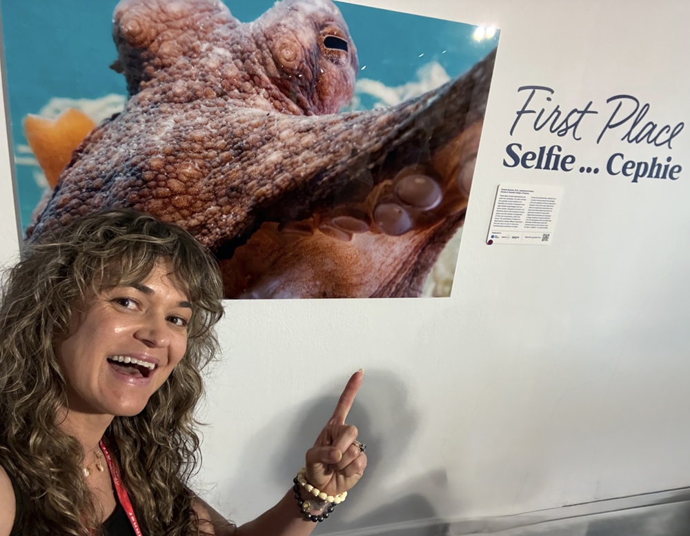
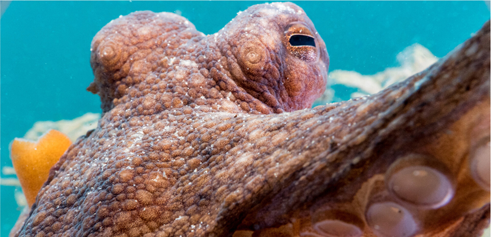
Dr. Chelsea Bennice, Research Fellow at the FAU Marine Lab and Glenn W. & Cornelia T. Bailey Marine SEA Scholar Program leader, won the first-place prize for her photo “Selfie…Cephie.” The image shows a curious cephalopod (an octopus) holding the camera with one of its flexible arms, seemingly taking a ‘selfie.’
“It’s an amazing feeling to be a scientist and a science communicator who captivated the audience through photography and storytelling,” Bennice shares. “Octopuses are having their moment! They are truly in the spotlight at this Art of Science Gallery for the community to see their beauty and how important they are for scientific discovery.”
Bennice studies the arm flexibility and behavior of octopuses. Their eight muscular (and boneless!) arms bend, shorten, elongate, and twist in all directions to create a complex behavioral repertoire crucial for survival. Bennice explains, “They are quite the impressive multitasker.” Bennice’s incredible photo is not just a part of her research. She tells us, “It wasn’t until I walked through the gallery’s doors on opening night and saw my image on a large acrylic canvas that I realized: ‘Wow, this is art.’”
|
It’s a selfie of a selfie as first-place photographer, scientist, and science communicator extraordinaire, Dr. Chelsea Bennice, snaps her own photo with her first-place submission: “Selfie…Cephie” – a play on the group of animals octopuses belong to, cephalopods. |
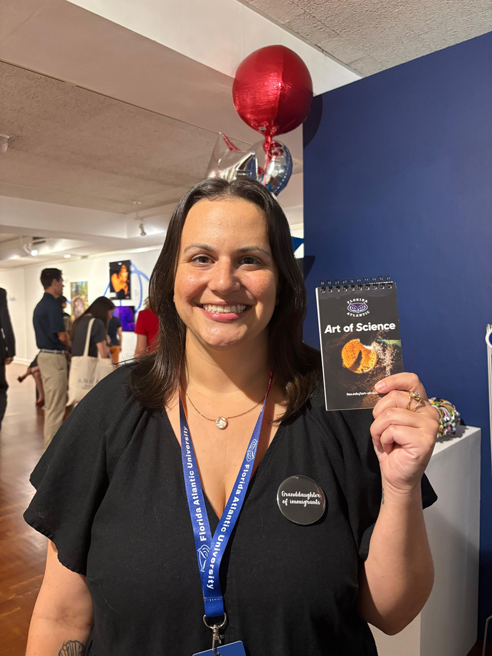
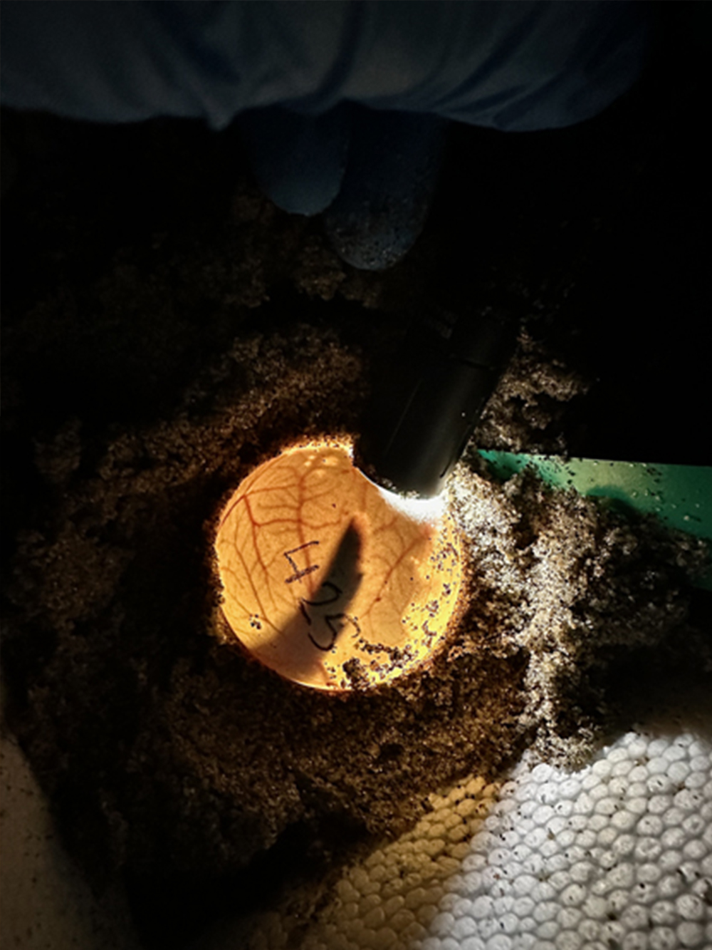
Alongside Bennice, FAU Marine Lab’s Gabby Carvajal also secured a major accomplishment, placing second in the Art of Science contest. Carvajal is a Ph.D. Candidate in Dr. Wyneken’s lab and a GW&CTB Marine SEA Scholar. Her photograph “Sea Turtle Beginning” gives a rare glimpse inside the embryonic development of a loggerhead sea turtle. Carvajal’s research involves incubating sea turtle eggs at controlled temperatures to investigate temperature-dependent sex determination. A technique called “candling,” in which a flashlight is held up against an egg in the dark, allows her to monitor the growing embryo and the intricate network of blood vessels that sustain its growth.
“It feels great to win second place,” Carvajal expressed. “It’s especially rewarding to see something I find so interesting resonate with a broader audience.” She was especially glad that the image so clearly showed the flipper outline of the embryo, which could “feel more recognizable" to the public audience. “What might first look abstract becomes something much more meaningful, especially since you’re witnessing an early stage of life that’s usually never seen,” Carvajal remarked.
|
|
| Second-place winner, Gabby Carvajal, poses with a small notebook with her submission printed on the cover! Her photo shows how FAU Marine Lab scientists monitor sea turtle development in the lab by "candling" the eggs. With a flashlight, one can see development in action from the blood supply to the close-up of the flipper. | |
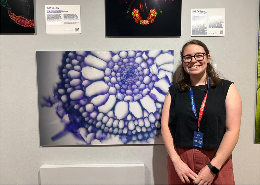
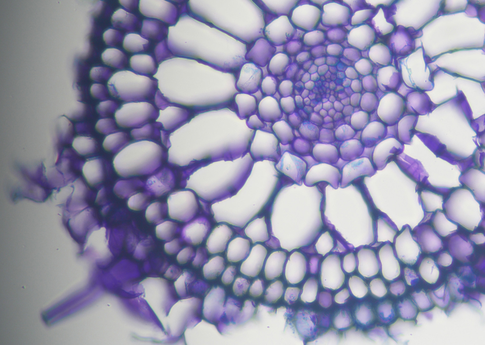
Alex Hoey, a former GW&CTB Marine SEA Scholar and current biology master’s student in Dr. Marguerite Koch’s lab, studies the gas exchange in seagrass roots present in the bottom of Florida Bay. Hoey earned honorable mention accolades and a place on the wall in the Art of Science Gallery for her image revealing the microscopic seagrass “oxygen highways” known as “aerenchyma” (ah-rank-ah-ma). Like plants on land, seagrasses create oxygen through photosynthesis; the aerenchyma in Hoey’s picture transports that oxygen from the leaves to the roots. Oxygen is often scarce in the sediment surrounding seagrasses, so these microscopic airspace tissues are vital for seagrass survival – and the survival of the many creatures that depend upon and reside within the seagrass ecosystem.
Photos like Hoey’s serve as a bridge between scientists and our broader community. Those photos are “…visually representing the methods or results of dynamic studies which allows for the inclusion of non-scientists into the science world, benefiting both our broader society and the field of science,” said Hoey.
|
|
| Alex Hoey poses with her honorable mention submission depicting the microscopic airspace tissues that supply seagrass roots with oxygen. | |
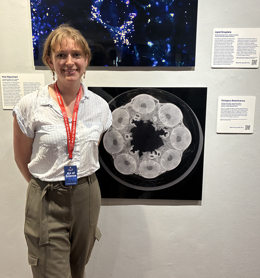
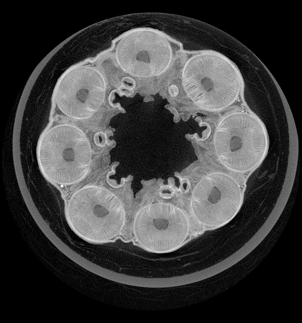
Natasha Ahrweiler, a biology master’s student working with Drs. Bennice and Randy Brooks, also had a submission earn honorable mention accolades in the Art of Science Gallery. Her picture, named “Octopus americanus,” depicts a Micro-CT cross-sectional image directly below the head of the octopus that shows the crown of eight arms. Using this non-invasive method, the internal morphology of the eight arms and each arm’s nerve cord remain intact and become visible.
“Micro-CT lets us get a glimpse of the internal structures of an animal as they are, without altering the specimen,” Ahrweiler said. “We get these beautiful images, and we get thorough data on size, layout, and composition of structures inside an animal, while also preserving the specimen for future use in other studies.”
“To me, visuals are imperative to understanding science,” Ahrweiler told us. “I think they engage our brain in a special way and can really make complex scientific concepts click into place.”
Rather than just viewing these visuals as data, Ahrweiler believes that science can truly be art: “All that knowledge you build as a scientist isn't just a block of text from a textbook – it's the actual, tangible makeup of the world around us.”
|
|
| Natasha Ahrweiler poses with her honorable mention submission depicting a micro-CT image cross-section directly below the head of an octopus. The unaltered crown of eight arms and their corresponding nerve cords are visible. | |
Our scientists were able to admire their own hard work and artistic prowess as their images were displayed, not just on a computer screen or as a figure in a peer-reviewed article, but in a true gallery setting. The work of Bennice, Carvajal, Hoey, Ahrweiler, and other FAU scientists captivated an audience and shared the beauty of the subjects of their research that often goes overlooked. You can still admire the Art of Science images and learn about the research they depict on the virtual Art of Science Guided Tour.